2022年第一期

6-OHDA诱导帕金森病模型小鼠多巴胺能神经元坏死性凋亡的可能机制

日期:2023-11-22

摘要

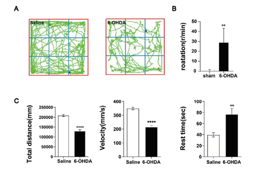
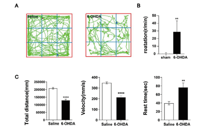
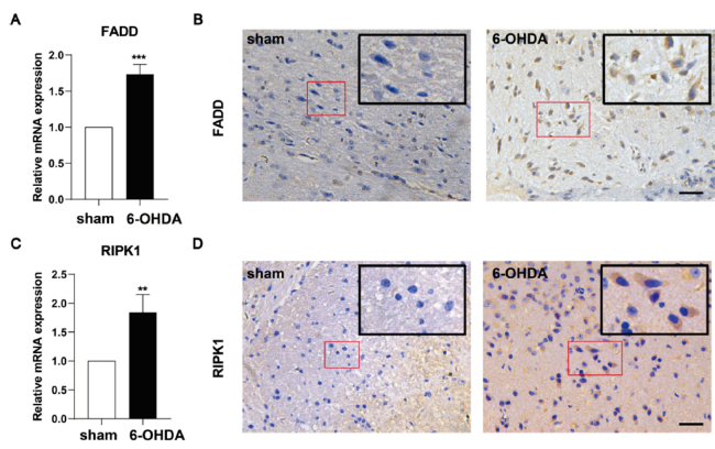
目的: 探讨6-羟基多巴胺氢溴酸盐(6-hydroxydopamine,6-OHDA)诱导帕金森病(parkinson's disease,PD)模型小鼠多巴胺能神经元坏死性凋亡的细胞机制。方法: 将含0.02% 抗坏血酸和0.9% 生理盐水的6-OHDA注入开颅后的8周龄、健康雄性C57/BL小鼠的单侧黑质(substantia nigra,SN) 区域构建PD小鼠模型(模型组),用APO旋尾实验和旷场实验验证PD小鼠模型的构建情况;取模型组小鼠脑组织进行免疫组化(immunohistochemistry,IHC)检测多巴胺能神经元(dopaminergic neurones,DA)标记物酪氨酸羟化酶(tyrosine hydroxylase, TH)恢复水平;通过qPCR和IHC检测模型组小鼠坏死性凋亡相关基因(FADD,RIPK1,TRADD)mRNA和蛋白表达水平。以左脑黑质区域只注入含0.02%抗坏血酸的0.9%生理盐水的小鼠作为正常对照组(假手术组)。结果: 与对照组比较,6-OHDA组小鼠的旋转次数明显增加,跑动距离和平均速度明显减少(P < 0.05),静止时间则明显增加(P < 0.05);6-OHDA组小鼠纹状体区域(STR)和黑质区域(SN)的TH阳性表达均明显降低(< 0.01),其中STR中的致密神经树突纤维明显减少,SN中的DA神经元大量丢失,仅可见少量多巴胺能神经元细胞存留,残留的阳性神经元细胞胞体小,突起短;6-OHDA组小鼠RIPK1、FADD的基因和蛋白表达水平均明显增加。结论: 6-OHDA可促进DA能神经元变性死亡,其可能机制是通过促进FADD-RIPK1坏死性凋亡信号通路。

Abstract

Objective: To investigate the cellular mechanism of 6-hydroxydopamine hydrobromide (6-OHDA) induced necrotic apoptosis of dopaminergic neurons in a parkinson's disease (PD) model mouse.Method: The model of Parkinson's disease was constructed by injecting 6-OHDA containing 0.02% ascorbic acid and 0.9% saline into the unilateral substantia nigra (SN) region of 8-week-old, healthy male C57/BL mouse after craniotomy, and the construction of the PD mouse model was verified by the APO spin tail test and the open field test. Brain tissues from model mouse were subjected to immunohistochemistry (IHC) to detect the recovery of dopaminergic neurones (DA) marker tyrosine hydroxylase (TH); the expression levels of FADD, RIPK1 and TRADD were detected by qPCR and IHC. The mRNA and protein expression levels of necrotizing apoptosis-related genes (FADD,RIPK1,TRADD) were measured by qPCR and IHC. A group of mouse injected with only 0.9% saline containing 0.02% ascorbic acid in the nigrostriatal region of the left brain was used as the normal control group. Result: Compared with the control group, mouse in the 6-OHDA group showed a significant increase in the number of rotations, a significant decrease in running distance and mean speed (P < 0.05), and a significant increase in resting time (P < 0.05). The positive expression of TH was significantly reduced in both striatal region (STR) and substantia nigra (SN) of mouse in the 6-OHDA group (P < 0.01), where dense nerve dendritic fibers were significantly reduced in STR and a large number of DA neurons were lost in SN, and only a few dopaminergic neuronal cells were seen to survive, with small cytosomes and short protrusions in the remaining positive neuronal cells. The gene and protein expression levels of RIPK1 and FADD were significantly increased in the 6-OHDA group of mouse. Conclusion: 6-OHDA promotes the degenerative death of Dopaminergic neurons by a possible mechanism that promotes the FADD-RIPK1 necroptosis signaling pathway.

关键词

帕金森病 / 坏死性凋亡 / 6-羟基多巴胺氢溴酸盐 / FADD / RIPK1

Key words

Parkinson's disease / Necrotic apoptosis / 6-OHDA / FADD / RIPK1

引用本文

徐志锋 练昌林 彭玲梅 赖文杰 周思捷 王玉凯6-OHDA诱导帕金森病模型小鼠多巴胺能神经元坏死性凋亡的可能机制. 阿尔茨海默病及相关病杂志. 2022, 5(1): 11-15 https://doi.org/10.3969/j.issn.2096-5516.2022.01.002
XU Zhi-feng LIAN Chang-lin PENG Ling-mei LAI Wen-jie ZHOU Si-jie WANG Yu-kaiStudy of the necrotic apoptosis effect in 6-OHDA-induced PD model mouse. Chinese Journal of Alzheimer's Disease and Related Disorders. 2022, 5(1): 11-15 https://doi.org/10.3969/j.issn.2096-5516.2022.01.002
 
帕金森病(parkinson's disease,PD)是目前人类第二常见的神经变性疾病,其发病率与患病率随年龄的增长而逐步上升,具有高病残率及低治愈率等特点,严重影响患者日常生活[1]。PD的主要病理改变是黑质多巴胺能神经元(DA)的变性死亡[2],但目前尚不清楚引起黑质多巴胺能神经元变性死亡的机制。传统观点认为在生理病理过程中,凋亡是唯一的程序性细胞死亡途径,而坏死则被认为是一种不可调控的过程[3]。随着对细胞死亡机制的不断深入研究,发现坏死并不是纯粹不受调控的、被动的细胞死亡方式,在某些情况下,是受一系列信号分子调控的、有序的过程[4]。近年来,坏死性凋亡在神经变性疾病发生发展中受到了越来越多的研究者的关注,有望为治疗这些疾病的药物开发提供潜在新靶点[5,6]。有研究报道,necrostatin-1可以有效抑制6-OHDA诱导的PC12细胞死亡[7],提示坏死性凋亡可能参与了6-OHDA对细胞的毒性作用。本研究旨探讨6-OHDA通过调控FADD-RIPK1坏死性凋亡信号通路促进PD模型小鼠DA能神经元变性死亡的可能机制。

1 材料和方法

1.1 实验动物

健康雄性C57/BL小鼠,8周龄,体重20~22 g,20只,购自广东省动物中心。

1.2 主要试剂及仪器

6-OHDA(含0.02% 抗坏血酸与0.9%生理盐水,5mg/mL,Sigma-Aldrich公司),SP试剂盒(SP-9001,ZSGB-BIO公司),SYBR Premix Ex Taq II(2×)试剂盒(TaKaRa公司),5×PrimeScript RT Master Mix试剂盒(Takara公司),明场显微镜(型号 DM2500;Leica公司)。

1.3 PD模型小鼠的制备及分组

取10只C57/BL小鼠作为模型组小鼠行开颅手,术前,小鼠禁食24 h,自由饮水,腹腔注射适量三溴乙醇(sigma) ;小鼠开颅后,将其固定于脑立体定位仪,前、后囟保持在同一水平面上。参照《小鼠脑立体定位图谱》[8],将含0.02 % 抗坏血酸和0.9%生理盐水的6-OHDA注入小鼠左脑黑质区域(substantia nigra,SN)(AP, -3 mm;ML, -1.3 mm;DV, -4.7 mm),以0.5 μL/min速度注射3 μL,注射完毕后留针5 min,缓慢退针;缝合伤口并敷青霉素以预防感染。同时取10只C57/BL小鼠作为正常对照组(假手术组),其小鼠左脑黑质区只注入3 μL含0.02 %抗坏血酸的0.9 %生理盐水。

1.4 模型组小鼠行为学检测

PD模型组小鼠手术3周后,腹腔注入APO 0.5 mg/kg诱发其旋转,连续观察30 min,如平均旋转次数 >7 r/min,即视所建帕金森病小鼠模型成立[9]

1.5 小鼠自主活动能力的检测

采用旷场试验(open field test,OFT) [10]。旷场实验箱高40 cm,边长100 cm,内壁不透明,正上方1.5 m处架一数码摄像头,其视野可覆盖整个旷场内部。实验前,将小鼠置于旷场箱中适应5 min,然后开始记录,共记录10 min。整个测试过程中应保持环境安静,测试结束后采用系统自带软件对2组每只小鼠跑动距离、行走速度以及静止时间等所记录的视频进行分析。

1.6 免疫组化

用三溴乙醇对小鼠实施安乐死,将4% 多聚甲醛(yong jin biotech) 经心脏灌注。 取小鼠脑组织,固定,制作厚4 µm(SN 和 STR 区域)石蜡切片,每只小鼠共制作6个冠状切片;参照SP试剂盒说明书处理脑组织切片, 一抗为抗酪氨酸羟化酶 (1:500)。用明场显微镜(型号 DM2500;Leica)和 ImageJ 软件(美国国立卫生研究院)测量 小鼠SN和 STR 区中 TH+ 神经元密度。

1.7 qPCR

用Trizol 提取试剂盒提取小鼠脑组织标本中总 RNA。由英潍捷基公司设计并合成了引物(见表1)。参照5×PrimeScript RT Master Mix试剂盒(Takara公司产品)说明书,将RNA 反转录为 cDNA。同时参照 SYBR Premix Ex Taq II(2×)试剂盒说明书,反应溶液用于荧光定量 PCR。荧光定量PCR 于 Roche LightCycler 480系统中进行。以甘油醛磷酸脱氢酶(GAPDH)作为 FADD和RIPK1的内部参数,检测FADD和RIPK1的表达水平。靶基因相对转录水平通过 2-△△Ct 法计算。
表1 qPCR引物

Tab.1 qPCR primers

引物序列信息(5'-3')
FADDGCGCCGACACGATCTACTG

TTACCCGCTCACTCAGACTTC
RIPK1GCACCCGAACACCTGAATGA

CGAACTGCTCAGTACAGATGACA
GAPDHAGGTCGGTGTGAACGGATTTG

GGGGTCGTTGATGGCAACA

2 结果

2.1 6-OHDA对PD模型组小鼠行为学的影响

结果显示,与对照组小鼠比较,模型组小鼠的跑动距离及平均速度明显减少(P < 0.05),静止时间明显增加(P < 0.05);同时,6-OHDA组小鼠活动范围较对照组小鼠明显杂乱无章,提示PD模型组小鼠的探索能力下降,即6-OHDA诱发了PD模型组小鼠的运动功能障碍,见图1。
图1 6-OHDA对PD模型组小鼠的行为学影响

Fig.1 Behavioral changes of PD mouse in 6-OHDA group

Note: Figure A:The two groups of mouse were evaluated for their voluntary motor function and exploratory function by the open field test, and it was seen that the mouse in the 6-OHDA group had a smaller range of motion and a more chaotic trajectory compared to the control group. Figure B:By the APO tail rotation experiment, the average number of rotations of the 6-OH group mouse were all more than 7r/min, and it was clear that the 6-OHDA mouse were successfully modeled. Figure C:In the open field test, there were significant differences in running distance, speed and resting time between the two groups of mouse. The total active distance and average moving speed of mouse in the 6-OHDA group were significantly reduced and the resting time was significantly increased.(* p < 0.05,**p < 0.001,*** p < 0.000 5,****p < 0.000 1)

Full size|PPT slide

2.2 6-OHDA对模型组小鼠DA能神经元变性的影响

与对照组比较,模型组小鼠的纹状体区域(STR)和黑质区域(SN)的TH表达均显著减少(P < 0.01),其中STR中的致密神经树突纤维明显减少,SN中的DA神经元大量丢失,仅可见少量多巴胺能神经元细胞存留,残留的阳性神经元细胞胞体小,突起短,其结果提示6-OHDA能促进PD小鼠DA能神经元神经变性死亡,从而引起PD小鼠运动功能障碍,见图2。
图2 6-OHDA促进DA能神经元变性死亡

Fig.2 6-OHDA promotes the degeneration and death of dopaminergic neurons

Note: FigureA-B:The positive expression of DA in the SN and STR of mouse was observed by IHC, and the DA in the NS in clusters and in the STR in dots could hardly be observed in the 6-OHDA group of mouse;Bar=200 μm

Full size|PPT slide

2.3 6-OHDA对 FADD 和 RIPK1 表达的影响

qPCR结果显示,与对照组小鼠比较,模型组小鼠的FADD、RIPK1基因表达及SN区域中的FADD阳性表达水平均明显增加;模型组中小鼠RIPK1的表达水平也明显增加,其主要分布于多巴胺能神经元细胞质中,上述结果提示6-OHDA能够诱发DA能神经元变性死亡,其可能机制是通过上调FADD-RIPK1坏死性凋亡信号通路,见图3。
图3 6-OHDA对 FADD 和 RIPK1 表达的影响

Fig.3 Influence of 6-OHDA on the expression of FADD and RIPK1

Note: FigureA,C:The expression of FADD and RIPK1 was detected by qPCR, and the histogram suggested that the expression levels of FADD and RIPK1 genes were significantly elevated in 6-OHDA. FigureB,D:The intracellular expression of FADD and RIPK1 was detected by IHC in two groups of mouse, and a significant increase in the expression of FADD and RIPK1 was seen in the brain tissue of mouse in the 6-OHDA group.(* p < 0.05,** p < 0.001,*** p < 0.000 5);Bar=100 μm

Full size|PPT slide

3 讨论

帕金森病作为人类第二常见的神经变性疾病,给社会以及患者家庭带来巨大的精神和经济负担[11]。然而,目前帕金森病的发病机制尚未明确,近年来研究者们提出,细胞坏死性凋亡可能在中枢神经系统退行性疾病中发挥着重要的作用[5。6-羟基多巴胺氢溴酸盐(6-OHDA)诱导的PD小鼠模型是PD研究领域的经典模型,可模拟类似于PD的许多病理特征和运动缺陷,使其成为探讨PD发病机制的常用模型[12]。坏死性凋亡是近年来发现的一种新的细胞死亡方式,是由死亡受体及其配体相连接而诱导激活,包括肿瘤坏死因子受体(TNF receptor,TNFR)1和2[13]。一些研究表明,坏死性凋亡与神经变性疾病的发生发展有着密切关系[14,15]。尽管有文献报道提示坏死性凋亡与PD体外模型有联系,但是关于帕金森病的发病病因与坏死性凋亡之间的关系仍有待阐明。有研究发现在6-OHDA处理前1h将necrostatin-1加入PC12细胞中可以有效抑制6-OHDA诱导的细胞死亡[7],提示坏死性凋亡可能参与了6-OHDA对细胞的毒性作用,然而目前鲜有研究者开展相关的动物实验。也有研究者提出坏死性凋亡参与了PD小鼠的发病过程[16],但对于具体的发病机制尚未进行进一步的探究。本研究则着重于探讨6-OHDA通过FADD-RIPK1坏死性凋亡信号通路轴促进DA能神经元变性死亡。
本研究结果显示,与对照组小鼠比较,6-OHDA可导致模型组小鼠STR和SN区域中DA能神经元变性死亡,明显减少PD模型组小鼠的自主运动能力以及探索能力。
细胞坏死性凋亡是一种通过破坏坏死细胞并释放细胞内成分来触发先天免疫反应的坏死。研究提示,细胞坏死性凋亡在帕金森病的发病中可能起重要的作用[17]。FADD是Roy[18]于1995年筛选出来的一个与细胞内Fas因子作用的蛋白,称为Fas相关死亡域蛋白(fas-associated death domain),坏死性凋亡中FADD的募集,可以介导半胱氨酸酶8(caspase 8)的激活,进而激活凋亡信号通路[19]。而受体相互作用蛋白激酶1 (receptor-interacting protein kinase 1, RIPK 1)是细胞凋亡和坏死以及炎症通路中的一个关键调节因子,可以通过与其他蛋白形成坏死体激活坏死性凋亡通路。本研究分别进行了IHC和qPCR实验检测,结果显示,与对照组小鼠比较,FADD及RIPK1在6-OHDA模型组小鼠的SN区域中蛋白表达明显增加,SN区域的FADD及RIPK1基因表达水平也明显上升,故本研究结果提示6-OHDA损伤DA能神经元可能与坏死性凋亡相关。
在本研究基础上,下一步我们将利用程序性凋亡相关抑制剂进一步验证6-OHDA通过坏死性凋亡途径促进DA能神经元变性死亡,并通过体外实验进一步探索6-OHDA促进DA能神经元坏死性凋亡的机制。

参考文献

[1]
Tysnes O B, Storstein A. Epidemiology of Parkinson's disease[J]. J Neural Transm (Vienna)2017124(8): 901-905.
[2]
Braak H, Tredici K D, Rüb U, et al. Staging of brain pathology related to sporadic Parkinson's disease[J]. Neurobiology of aging200324(2):197-211.
[3]
伏媛. 程序性坏死研究进展[J]. 国际病理科学与临床杂志201232(1):50-54.
[4]
Galluzzi L, Vitale I, Aaronson S A, et al. Molecular mechanisms of cell death: recommendations of the Nomenclature Committee on Cell Death 2018[J]. Cell Death Differ201825(3):486-541.
[5]
Dionisio P A, Amaral J D, Rodrigues C. Molecular mechanisms of necroptosis and relevance for neurodegenerative diseases[J]. Int Rev Cell Mol Biol2020353:31-82.
[6]
Salvadores N, Court F A. The necroptosis pathway and its role in age-related neurodegenerative diseases: will it open up new therapeutic avenues in the next decade?[J]. Expert Opin Ther Targets202024(7): 679-693.
[7]
Wu J R, Wang J, Zhou S K, et al. Necrostatin-1 protection of dopaminergic neurons[J]. Neural Regen Res201510(7):1120-1124.
[8]
Cirmi S, Maugeri A, Lombardo G E, et al. A Flavonoid-Rich Extract of Mandarin Juice Counteracts 6-OHDA-Induced Oxidative Stress in SH-SY5Y Cells and Modulates Parkinson-Related Genes[J]. Antioxidants (Basel)202110(4): 539.
[9]
Pan Q, Zhang W, Wang J, et al. Impaired voluntary wheel running behavior in the unilateral 6-hydroxydopamine rat model of Parkinson's disease[J]. J Korean Neurosurg Soc201557(2):82-87.
[10]
Lian C, Huang Q, Zhong X, et al. Pentraxin 3 secreted by human adipose-derived stem cells promotes dopaminergic neuron repair in Parkinson's disease via the inhibition of apoptosis[J]. FASEB J202135(7):e21748.
[11]
Global, regional, and national burden of neurological disorders, 1990-2016: a systematic analysis for the Global Burden of Disease Study 2016[J]. Lancet Neurol201918(5):459-480.
[12]
杨东明, 杨利峰, 赵德明, 等. 帕金森病动物模型的研究进展[J]. 中国实验动物学报202028(3):397-404.
[13]
Laster S M, Wood J G, Gooding L R. Tumor necrosis factor can induce both apoptic and necrotic forms of cell lysis[J]. J Immunol1988141(8): 2629-2634.
[14]
杨海玉, 刘勇. 坏死性凋亡与中枢神经系统退行性疾病的研究进展[J]. 中国神经免疫学和神经病学杂志201724(2):143-146.
[15]
Jantas D, Greda A, Golda S, et al. Neuroprotective effects of metabotropic glutamate receptor group II and III activators against MPP(+)-induced cell death in human neuroblastoma SH-SY5Y cells: the impact of cell differentiation state[J]. Neuropharmacology201483:36-53.
[16]
尹秀华. PD模型大鼠神经细胞程序性凋亡通路研究及坏死性凋亡初探[D]. 苏州大学免疫学, 2014.
[17]
杨璇, 杨兵, 张翼, 等. 帕金森病中多巴胺能神经元的程序性细胞死亡机制[J]. 神经损伤与功能重建201813(6):296-299.
[18]
Roy U. Structural Characterizations of the Fas Receptor and the Fas-Associated Protein with Death Domain Interactions[J]. Protein J201635(1):51-60.
[19]
Hartmann A, Mouatt-Prigent A, Faucheux B A, et al. FADD: A link between TNF family receptors and caspases in Parkinson's disease[J]. Neurology200258(2):308-310.